Overview
The main area of the brain involved in Huntington's disease (HD) is the basal ganglia, which is made up of the caudate nucleus, the putamen, the globus pallidus (interna and externa), and the subthalamic nuclei (STN); the substantia nigra (SNC and SNR) of the midbrain is also connected to the basal ganglia. One of the basal ganglia's main functions is in the control and processing of movement.
The basal ganglia lie deep within the two hemispheres, and are responsible for initiation of movement.
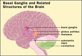
Image of the Basal Ganglia. Image courtesy of commons.wikimedia.org/wiki/File:Brain_structure.gif
Neurotransmitters in the Basal Ganglia
GABA
GABA is the main inhibitory neurotransmitter in the CNS. It acts at a ligand-gated anion-selective channel; when GABA binds it allows chloride ions to enter the cell, decreasing the membrane potential and therefore hyperpolarising the cell.
GABA is also strongly implicated in anxiety.
Glutamate
Glutamate is the main excitatory neurotransmitter of the CNS. It also acts at ligand-gated ion channels; the NMDA receptors, the AMPA receptors and the kainate receptors, as well as the metabotropic mGluR receptor (a GPCR). Glutamate binding allows calcium to enter the cell and therefore depolarise it, generating action potentials.
Glutamate receptors are also implicated in learning and memory, hence their role in cognition.
Dopamine
Dopamine, the precursor for noradrenaline, acts via D2 receptors in this pathway. It inhibits cells of the striatum, which therefore decreases the inhibition from the striatum onto the globus pallidus and therefore increases action potential firing from the thalamus. The dopamine receptor is a GPCR.
Dopamine is also implicated in learning, memory and emotions. It is also affected in Parkinson's disease.
The Basal Ganglia network
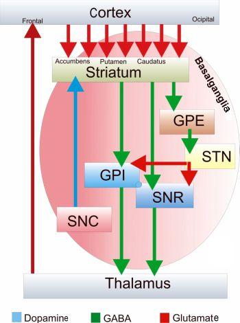
There are several pathways of excitation and inhibition, as shown in the above diagram.
Image showing the pathways of the Basal Ganglia.Image adapted courtesy of commons.wikimedia.org/wiki/File:Basal-ganglia-2.jpg
At rest, neurons of the globus pallidus are spontaneously active and inhibit the thalamic nuclei (the VL) through GABA release.
When the cortex is activated, so are the neurons of the putamen, which then inhibit the globus pallidus (through the release of GABA); this therefore removes the inhibition of the thalamus from the GPI, meaning the thalamus is now active. This produces a positive feedback loop, as the thalamus then further excites the supplementary motor area (SMA) of the cortex through glutamatergic pathways, and allows movement to be generated.
A simplified version of the motor loop:
Excites Inhibits Releases inhibition of Boosts activity of
Cortex ------------> Striatum --------------> Globus Pallidus ------------------> VL (Thalamus) -----------------> Cortex (SMA)
Together, the caudate nucleus and the putamen form the striatum, and the globus pallidus and putamen form the lentiform nucleus.
Huntington's disease
HD leads to a loss of neurons, most notably in the caudate nucleus, putamen and globus pallidus; all three normally produce inhibitory effects on the substantia nigra. The loss of neurons therefore leads to a loss of inhibition to the thalamus, resulting in spontaneous uncontrollable movement known as chorea.
As well as being responsible for the motor aspects of HD, the basal ganglia are also important in the linguistic and cognitive aspects. The disease is not only characterised by excessive movement, but also excessive rule use in language. Language tests demonstrate that sufferers multiply suffix some verbs, for example look-eded instead of look-ed, as the suffixing rule is over-active.